An article published in the “Scientific Reports” journal reports a 3D reconstruction of some of the oldest known dinosaur embryos, which belong to the species Massospondylus carinatus. Kimberley “Kimi” Chapelle, Vincent Fernandez, and Jonah Choiniere subjected the fossils to a very special CT-scan, an exam that was conducted at the European Synchrotron Radiation Facility (ESRF) in Grenoble, France, using an 844-meter-long particle accelerator. The result is that the eggs containing those embryos fossilized when they were at about 60% of their development, which is similar to that of some of the closest relatives of the dinosaurs existing today: crocodiles, chickens, turtles and lizards.
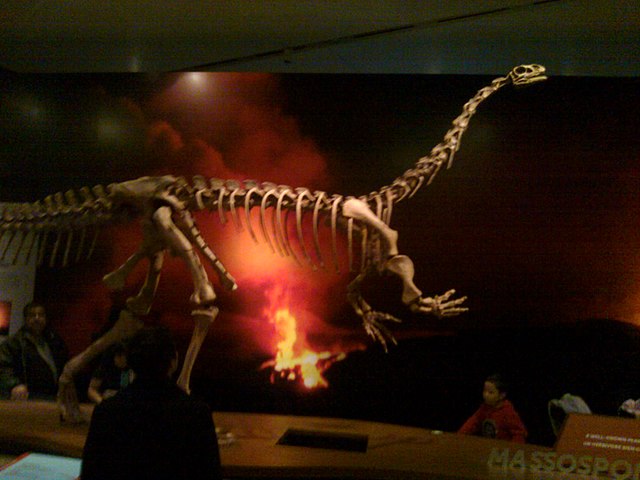
A clutch with seven fossil eggs of Massospondylus carinatus (photo of skeleton reconstruction ©Dinalfos5), a herbivorous dinosaur that lived in the Triassic and Jurassic periods, was discovered in 1976 in the Elliot Formation, in the Golden Gate Highlands National Park, South Africa. Two partially exposed embryos made it possible to identify them as dinosaurs, making the eggs among the oldest known among dinosaur eggs. The problem so far was in the difficulty of studying tiny and very fragile fossils, characteristics that made them of little use. For example, paleontologists believed that those Massospondylus carinatus’ eggs started fossilizing just before hatching but it was really difficult to verify that theory.
New technologies are making various advances in the field of paleontology, for example with non-destructive tests. Versions of the CT-scan adapted to the study of fossils now allow to create 3D reproductions and study them in detail. In the case of Massospondylus carinatus eggs, the CT-scan is very different from the one performed in a hospital on humans because the resolution that reaches the levels of the single bone cell was achieved only thanks to a particle accelerator.
In common CT-scans, radiation must be dosed in order to avoid damage to patients, in the case of fossils there’s no such problem. However, an in-depth examination such as the one carried out at the ESRF took a very long time to bring the results wanted. In fact, the scan was conducted in 2015, and it took about three years to process the data obtained at the University of Witwatersrand in South Africa. An adult Massospondylus carinatus could be 5-meter long but the skull of the embryos examined is only about 2 centimeters long. The amount of detail collected even for such small embryos took all that time to create the 3D model.
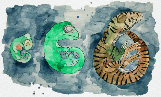
The study of the collected data offered various information on the embryos. The bones of the skull show that the embryos were at about 60% of the incubation period and not close to hatching, as previously thought. The top image (Courtesy Mélanie Saratori) shows a watercolor illustration of Massospondylus carinatus embryos at 17%, 60% and 100% of their incubation period.
The researchers also found that the embryos had two types of teeth. A set consisted of very simple triangular-shaped teeth that would have been reabsorbed or shed before hatching, as is the case today for crocodiles and geckos. The second set was very similar to that of adults and the embryos would have been born with those teeth. These were really tiny teeth, with a width between 0.4 and 0.7 millimeters.
One conclusion is that the development of these dinosaurs’ embryos was similar to that of their reptilian relatives. Further information may come from the continuation of the exams, which in the first phase were focused on the embryos’ skull.